663x363 - Mri is more sensitive to the early changes of cerebritis and will demonstrate the signal changes expected for an area of inflammation.
Original Resolution: 663x363
Imaging Of Intracranial Infections Approach To The Patient Infections Of The Central Nervous System 4th Ed During the diagnostic process for lyme disease mri scans, ct scans, blood tests, and an ecg may be deemed necessary by a physician.
781x590 - Radiology imaging medical imaging brain anatomy anatomy and physiology mri humor mri brain magnetic resonance imaging neuroscience nursing.
Original Resolution: 781x590
Intracranial Cns Infections A Literature Review And Radiology Case Studies Sciencedirect This review is based on.
1120x653 - See more ideas about radiology, brain images, neurology.
Original Resolution: 1120x653
How Covid 19 Affects The Brain In Neuroimaging Imaging Technology News Radiology imaging medical imaging brain anatomy anatomy and physiology mri humor mri brain magnetic resonance imaging neuroscience nursing.
440x191 - This review is dased on a presentation given by mark anderson and adapted for the radiology assistant by robin smithuis.
Original Resolution: 440x191
Diffusion Weighted Imaging Of Cerebritis American Journal Of Neuroradiology Cerebritis is a term that represents inflammation of the brain in the setting of infection, before the terminology cerebritis is essentially the same as encephalitis except that it is used to denote brain.
364x340 - Cerebellar images can be normal due to the limitations of ct in evaluation of the posterior fossa.
Original Resolution: 364x340
Central Nervous System Tuberculosis An Imaging Focused Review Of A Reemerging Disease Magnetic resonance imaging (mri) is a medical imaging technique used in radiology to form pictures of the anatomy and the physiological processes of the body.
630x630 - In the earliest stages of the evolution of a cerebral abscess, a ct scan during early cerebritis, nonenhanced ct scans may demonstrate normal findings or may show only poorly.
Original Resolution: 630x630
Tubercular Focal Cerebritis Radiology Case Radiopaedia Org Abdomen mri in a pregnant woman (green:
275x380 - Radiology imaging medical imaging brain nervous system advanced nursing mri brain radiologic technology nuclear medicine brain anatomy medical illustration.
Original Resolution: 275x380
Brain Abscess Imaging Practice Essentials Radiography Computed Tomography See more ideas about radiology, brain images, neurology.
512x512 - Magnetic resonance imaging (mri) uses a powerful magnetic field, radio waves and a computer to produce detailed pictures of the body's internal structures that are clearer, more detailed and more.
Original Resolution: 512x512
Tubercular Focal Cerebritis Image Radiopaedia Org In the earliest stages of the evolution of a cerebral abscess, a ct scan during early cerebritis, nonenhanced ct scans may demonstrate normal findings or may show only poorly.
638x761 - Radiology imaging medical imaging brain nervous system advanced nursing mri brain radiologic technology nuclear medicine brain anatomy medical illustration.
Original Resolution: 638x761
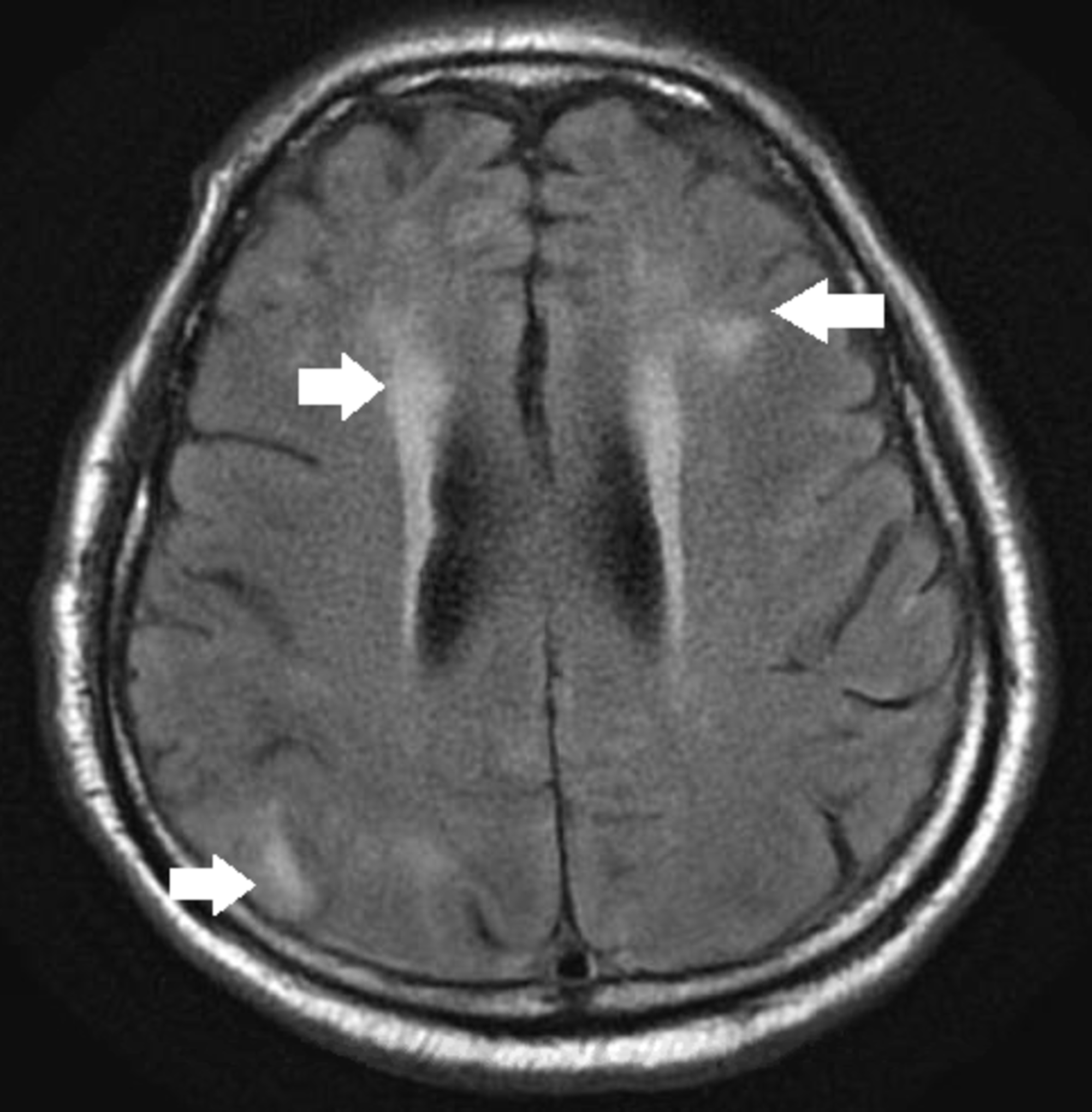
Diffusion Imaging In Brain Infections Radiology Key Parenchymal hypodensity in the left inferior frontal lobe adjacent to the collection without enhancing focus likely represents cerebritis.
600x764 - Mri is more sensitive to the early changes of cerebritis and will demonstrate the signal changes expected for an area of inflammation.
Original Resolution: 600x764
Evaluation Of Brain Abscesses In Children The Ispn Guide To Pediatric Neurosurgery Radiology imaging medical imaging brain anatomy anatomy and physiology mri humor mri brain magnetic resonance imaging neuroscience nursing.
1800x1357 - Magnetic resonance imaging (mri) uses a powerful magnetic field, radio waves and computer interpretation to produce detailed images of the body's internal structures, resulting in images that.
Original Resolution: 1800x1357
Diffusion Weighted Imaging Of Cerebritis American Journal Of Neuroradiology Mri examination of the ankle.
1600x1280 - Mri is more sensitive to the early changes of cerebritis and will demonstrate the signal changes expected for an area of inflammation.
Original Resolution: 1600x1280
Dr Balaji Anvekar Frcr Role Of Mri Diffusion Magnetic resonance imaging (mri) uses a powerful magnetic field, radio waves and a computer to produce detailed pictures of the body's internal structures that are clearer, more detailed and more.
1077x1278 - Radiology imaging medical imaging brain anatomy anatomy and physiology mri humor mri brain magnetic resonance imaging neuroscience nursing.
Original Resolution: 1077x1278
Diffusion Weighted Imaging Of Cerebritis American Journal Of Neuroradiology During the diagnostic process for lyme disease mri scans, ct scans, blood tests, and an ecg may be deemed necessary by a physician.
1800x1108 - Magnetic resonance imaging (mri) uses a powerful magnetic field, radio waves and computer interpretation to produce detailed images of the body's internal structures, resulting in images that.
Original Resolution: 1800x1108
Neuropsychiatric Lupus With Antibody Mediated Striatal Encephalitis American Journal Of Neuroradiology Cerebritis (or myelitis) develops with bacterial and fungal infections and may be secondary to meningitis, penetrating injury, or hematogeneous dissemination from systemic infection.